Discover the Power of Precision Imaging for Temporomandibular Joint (TMJ) Disorders
If you’re suffering from jaw pain, clicking sounds when chewing, headaches, or difficulty opening your mouth, a TMJ MRI (Temporomandibular Joint Magnetic Resonance Imaging) may be essential to pinpoint the root cause.
At MRI Dubai, we provide high-resolution TMJ MRI scans using the advanced 3 Tesla (3T) wide-bore MRI, which delivers superior image clarity and diagnostic accuracy.
What is a TMJ MRI?
A TMJ MRI is a specialized magnetic resonance imaging scan used to evaluate the temporomandibular joint, which connects your jawbone to your skull. This joint plays a crucial role in speaking, chewing, and facial expressions. An MRI is the gold standard imaging modality for TMJ disorders because it provides detailed views of soft tissues, cartilage, discs, ligaments, and joint fluid—all without the use of radiation.

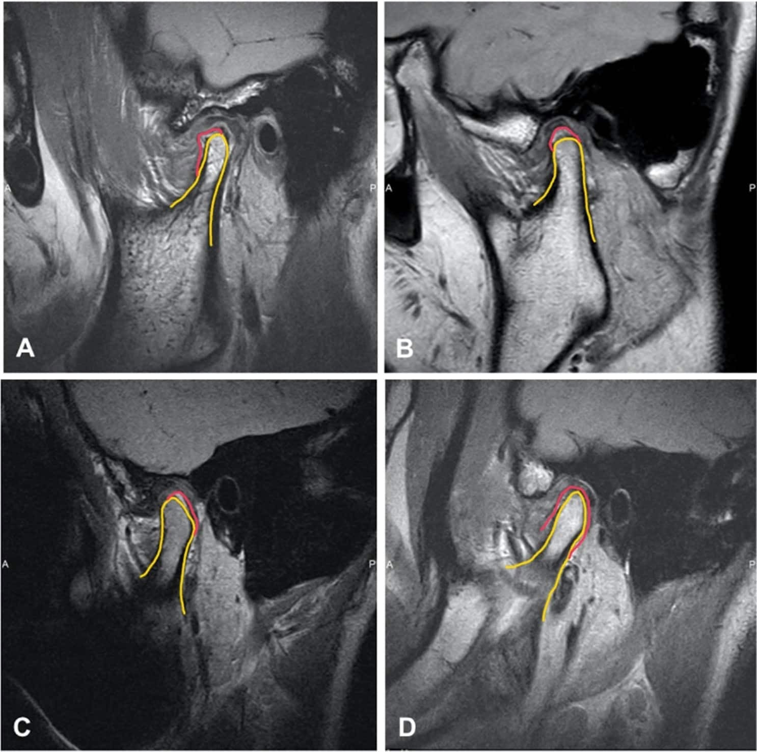
Magnetic resonance imaging (MRI) is one of the best diagnostic tools for identification of TMJ pathology, allowing evaluation of TMJ disc position, morphology, mobility, extent of joint degenerative changes, inflammation, and presence of connective tissue/autoimmune diseases.
There is special TMJ coil for this MRI. Generally, most pictures will be done with your mouth closed. However, the last sequence of pictures requires you to keep your mouth open, biting on a splint. Therefore , imaging will be captured with open mouth and closed mouth. Correspondingly, this allows visualization of the TMJ’s and surrounding structures in the open position. At the same time, your scan will take approximately 30 minutes.
Two position for TMJ MRI ( Open-Mouth and Closed-Mouth)
TMJ dysfunction is dynamic—it often involves movement and disc displacement that occurs during jaw opening or closing. Therefore, a comprehensive TMJ MRI must be performed in both closed-mouth and open-mouth positions to:
- Closed-Mouth Position: You do not need anything in your mouth. You will simply keep your teeth together in a natural, relaxed bite.
- Open-Mouth Position: To capture the jaw fully open, the technician will use a Bite Block or Mouth Prop.
- Bite Blocks: These are typically small, rubberized, or plastic blocks placed between your upper and lower teeth to hold the jaw open at a specific height.
Closed-Mouth TMJ MRI – Structural Assessment at Rest
The closed-mouth view captures the resting position of the temporomandibular joint. This position is essential for evaluating:
- Articular disc position at rest
- Joint space and disc alignment between the mandibular condyle and temporal bone
- Signs of disc displacement without reduction
- Bone marrow signal abnormalities, indicating early arthritis or inflammation
- Joint effusion (fluid accumulation)
- Degenerative changes like osteoarthritis or erosion of the condyle
- Posterior band of the disc and whether it sits at the 12 o’clock position (normal)
This image helps determine whether the disc is in the correct place when the jaw is closed, which is often where disc displacements first become evident.
Open-Mouth TMJ MRI – Dynamic Function and Disc Mobility
The open-mouth view is vital to assess how the TMJ functions during movement. This position reveals:
- Whether the displaced disc reduces (moves back into place) during jaw opening
- Anterior disc displacement with or without reduction
- Disc mobility and whether the movement is symmetrical on both sides
- Joint congruency and condyle translation (how the jaw bone moves forward)
- Dynamic function of ligaments and soft tissues
- Evaluation of joint locking or restricted motion
- Confirmation of mechanical dysfunction that may not be visible in the closed-mouth view
This functional imaging helps determine whether treatment should be conservative (splints, physiotherapy) or interventional (injections or surgery).
These two positions together provide a comprehensive, dynamic evaluation of the TMJ and are essential for accurate diagnosis and targeted treatment planning.