Understanding Filler MRI: A New Standard in Safety and Precision
Cosmetic filler shots in the face and butt are very common these days in Dubai. People use them for volume, shape, and a younger look, so they choose fillers often. The results can look very good, but many people feel unsure after the injection. Filler MRI in Dubai helps to find out these questions.
For example, some people do not know what type of filler is used. Also, some worry that the filler moves from the original place. In addition, some feel pain, swelling, or hard areas. Sometimes, they see uneven or irregular shapes.
Because of all these issue , many people now want to do Filler MRI which it is a safe way to check the filler. Filler MRI shows the filler type, and location of the filler . In addition , it shows if it needs treatment or not. So, some people want to remove the filler.
At MRI Dubai Center-Jumeirah, our 3 Tesla (3T) MRI system offers ultra high resolution imaging to evaluate facial and gluteal fillers with maximum accuracy . The procedure is non-invasive, radiation-free, and designed for patient comfort. 3 Tesla Filler MRI shows all side effects caused by filler shot.
Filler MRI with and without contrast is a gold standard exam. It shows the filler composition. Also , It shows the filler brand type, location, the enhancement pattern and any complications.
This information is very important for doctors. As, filler MRI result , helps doctors plan filler removal , treatment or correction.

Why Choose 3 Tesla MRI for Filler Evaluation?
3 Tesla MRI is Gold Standard for evaluating dermal fillers .Moreover, The 3 Tesla (3T) MRI scanner offers twice the magnetic field strength of traditional MRI systems, delivering superior image quality and faster scan times. We Recommend to combine MRI with high frequency ultrasound.
Unlike ultrasound or CT Scan , 3 Tesla MRI shows soft tissue very clearly. It helps doctors see the filler type, its location, and its effect on nearby tissue
Filler MRI is particularly effective for:
- filler composition (hyaluronic acid, silicone, polyacrylamide, biopolymer, etc.)
- Mapping filler distribution across muscle, fat, or subcutaneous tissue
- Detecting complications, such as inflammation, granulomas, infection, or migration
- Evaluate vascular activity or inflammatory reactions
- Identification of tiny filler deposits or small injection areas
- Clear differentiation between filler and natural fat
- Visualization of contrast uptake patterns for biological activity
- Precise mapping for safe surgical or non-surgical removal
Filler MRI With vs Without Contrast – What’s the Difference?
A Filler MRI without contrast shows the location and distribution of fillers .However, filler MRI with contrast, can also assess tissue activity. Therefore, both filler MRI with and without contrast shall be done. Like , it shows signs of inflammation, infection or artery changes.
At MRI Dubai, our standard protocol includes both without and with contrast imaging for maximum accuracy.
Types of Filler combination
A variety of temporary and permanent filler agents are available , including
Calcium hydroxylapatite: FDA- approval since December 2006 for the treatment of facial and wrinkles.
Collagen: Collagen fillers are based on naturally occurring protein derived from a variety of sources.
liquid silicone it is permanent agent . It is mainly for facial augmentation and treatment of acne scars for more than 50 years
polytetrafluoroethylene: It is for facial augmentation since the mid-1990s, particularly in the lips, nasolabial folds, and glabella
hyaluronic acid: It is acid–based gel fillers (Restylane, Perlane, Juvéderm). It is not permanent fillers and it use for both facial rejuvenation and butt.
poly-l-lactic acid: It is FDA approval since 2004 for treatment of facial lipoatrophy in patients with HIV, but it is also used for facial rejuvenation.
Classification of Filler Injections According to Anatomical Site
Selecting filler shot depends on the area of the body. Softer fillers like hyaluronic acid are commonly used for the face, especially for the lips, cheeks, and under-eye areas. They restore volume and smooth wrinkles. Body and buttock fillers use denser, longer-lasting materials such as poly-L-lactic acid or calcium hydroxyapatite to enhance shape and contour. Experts choose each filler type based on the depth, elasticity, and movement of the area for a perfect result.
Filler MRI for Buttocks (Gluteal Area)
Buttock filler enhance contour or add volume. However, not all fillers are suitable for deep soft tissue use.
Common fillers for the buttocks include hyaluronic acid fillers for immediate volume and contour, and collagen-stimulating fillers like Sculptra to promote natural collagen production over time. In many cases, patients are unaware of which material was injected—making MRI evaluation essential for proper management.
3T MRI can identify:
– The exact filler type – hyaluronic acid, silicone, biopolymer, or mixed substances
– The layer and spread – intramuscular, subcutaneous, or migrated filler
– Complications – abscess, fibrosis, granuloma, necrosis, or infection
– Contrast enhancement – indicating tissue inflammation or vascular response
Filler MRI for Face – Detailed Evaluation for Cheeks, Lips, Chin, and Jawline
Facial filler are among the most popular cosmetic treatments for both individuals . They are commonly used to define cheeks, lips, chin, jawline , face and eyes.
In the late 19th century, the first substance that used for facial fillers was paraffin. But it was soon discontinued because of serious side effects such as migration, embolization, and granuloma formation. Today, many safe and approved filler materials are available for facial rejuvenation. These modern fillers are popular because they provide natural aesthetic results without the need for invasive surgery.
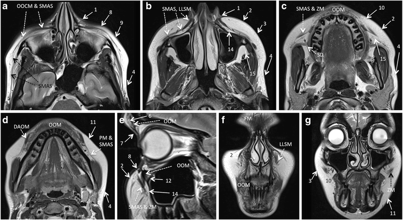
However, the wide variety of available products can make post treatment evaluation challenging. That’s where 3 Tesla MRI becomes invaluable. Patients with facial fillers often need imaging to check for complications such as infection, migration, overfilling, scarring, or foreign-body reactions.

Complications from filler injections can appear gradually over time. MRI with contrast detects problems such as:
– Filler migration beyond the intended area
– Inflammatory nodules or granulomas
– Abscesses or infection
– Fibrosis and encapsulation
– Tissue necrosis or vascular compromise
Early MRI evaluation allows timely, minimally invasive management—reducing the risk of long-term damage or disfigurement.
Patient Experience at MRI Dubai
At MRI Dubai, we focus on safety, comfort, and clarity. The Filler MRI is a completely non-invasive, radiation-free procedure.
What to Expect:
1. You’ll lie comfortably on the MRI table.
2. Non-contrast images are taken first.
3. A safe gadolinium contrast is injected intravenously.
4. The contrast scan follows immediately, providing enhanced images.
5. Our radiologist reviews both image sets and prepares a detailed report.
The total scan time takes around 45 minutes, depending on the area studied. You can return to normal activities right after the scan.
Conclusion: Advanced Filler MRI for Face and Body
MRI is mainly for individuals who have undergone filler injections in the buttocks or face and wish to evaluate, confirm, or remove them safely. Filler MRI with and without contrast offers the most advanced diagnostic insight available.
At MRI Dubai, our 3 Tesla MRI technology provides precise, high-resolution imaging to identify filler composition, size, brand, enhancement, and possible complication.
Book Your Filler MRI Today
If you have had filler injections in your face or buttocks and wish to assess or remove them safely, schedule your Filler MRI today.
Our 3 Tesla MRI system ensures unparalleled image quality, precise filler identification, and complete diagnostic confidence.
Website: www.mridubai.com
Contact: Book your appointment today . Take the next step toward safe and confident at MRI Dubai.
A Filler MRI is a specialized scan that shows the exact location and condition of cosmetic fillers in the face or body. It helps detect filler migration, lumps, or complications using high-resolution imaging.
A 3 Tesla MRI provides ultra-clear images — much sharper than standard MRI machines. It allows radiologists to accurately assess filler type, placement, and any possible reactions or inflammation.
Yes. MRI scans are completely safe and non-invasive. There’s no radiation involved, and our experienced radiology team ensures your comfort and safety throughout the scan.
Common areas include the face, lips, chin, and buttocks, and any filler region upon request.
A typical scan takes about 40-45 minutes, depending on the area being examined. You can resume normal activities immediately after.
You can book your scan directly at MRI Dubai in Jumeirah, and our specialists will guide you through the process
Key Takeaways
- Filler MRI uses advanced 3 Tesla technology to evaluate cosmetic fillers with high accuracy and safety.
- This non-invasive procedure helps identify filler types, locate them, and assess complications like migration or infection.
- Combining non-contrast and contrast MRI enhances diagnostic details, revealing tissue activity around fillers.
- Soft tissue contrast in MRI is superior to ultrasound or CT scans, making it ideal for filler evaluation in the face and body.
- Patients can comfortably undergo the procedure at MRI Dubai, with results aiding in informed treatment decisions.
Estimated reading time: 8 minutes
After struggling for months with complications from old fillers, I finally found answers at MRI Dubai. Their 3 Tesla filler MRI showed the exact location and specification of the filler, which allowed my doctor to plan the removal properly. I feel so relieved and grateful — truly professional service and very kind staff.
I did my filler MRI at MRI Dubai using their advanced 3 Tesla machine and I’m honestly very impressed. The report clearly showed exactly where the filler was placed and what type it was, which helped my doctor decide how to remove it safely. The whole process was fast, professional, and very reassuring. Highly recommended — thank you to the MRI Dubai team.
Very happy with my filler MRI at MRI Dubai 🙏 The 3 Tesla scan showed exactly what filler I had and where it was placed, so my doctor could remove it safely. Super clear report, fast service, and amazing team. Thank you!
I underwent a filler MRI at MRI Dubai using their 3 Tesla scanner, and the level of detail in the report was excellent. It clearly identified the filler type and distribution, which allowed my physician to plan targeted removal. Very professional service and high-quality imaging.